About Us
Experts in non-destructive 3D X-ray histology
Powered by the
μ-VIS X-ray Imaging Centre & Biomedical Imaging Unit

Located at the University Hospital Southampton the XRH Facility is a leading provider of 3D X-ray histology services, specialising in the imaging of formalin-fixed paraffin-embedded (FFPE) tissue samples.
We support both academic researchers and industrial partners, offering advanced solutions that drive innovation in biomedical and clinical transitional research, pharmaceutical technology and medical devices.
We are part of the University of Southampton’s μ-VIS X-ray Imaging Centre and the Biomedical Imaging Unit, and the EPSRC National Research Facility (NRF) for laboratory-based X-ray computed tomography, serving as the designated site for biomedical imaging.
Unique Capabilities
“Habitat”
Embedded within the University Hospital Southampton (UHS) Clinical Environment, operating alongside the Biomedical Imaging Unit (BIU), Histochemistry Research Facility, and the Biomedical Research Facility (BRF) we provide a synergistic ecosystem for high-impact research.
Custom Imaging Systems
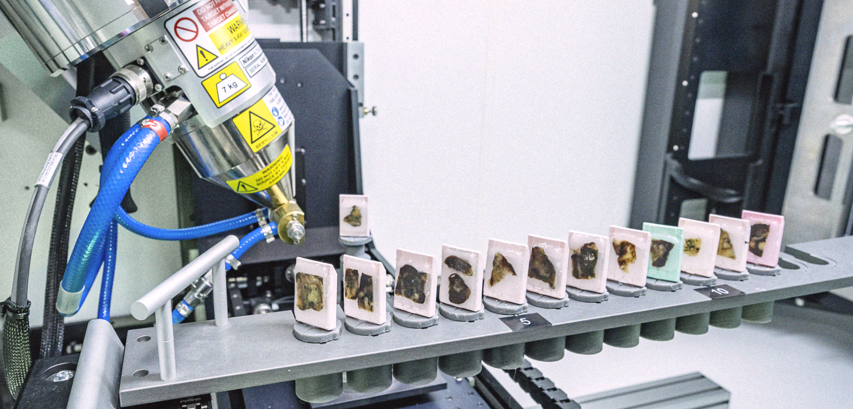
High-throughput imaging systems tailored for clinically relevant applications, designed to meet the specific demands of diverse research projects.
Correlative Imaging
Combining X-ray histology with conventional histology, immunohistochemistry, electron microscopy (SEM, SBFSEM, TEM), and spatial -omics.
Advanced Data Management
X-ray Histology Management System (XRHMS): In house developed fully functional and continuously evolving platform for managing histology samples, data, and metadata.

3D X-ray histology allows:
✓ Non-destructive, distortion-free 3D (volume) imaging of conventionally prepared FFPE tissue specimens
✓ Imaging of conventionally prepared formalin-fixed paraffin-embedded tissue
✓ Spatial resolution down to 5 μm
✓ Correlative 2D histology and immunohistochemistry and 3D visualisation
✓ Whole block imaging
✓ A multitude of 2D and 3D visualisation modes similar to the ones used in clinical radiology, including: multi-planar reconstruction (MPR); maximum intensity projections (MIP); interactive volume rendering; ‘on-the-fly’ arbitrary slicing
Our portfolio
3D X-ray Histology
We develop and deliver cutting-edge 3D X-ray histology imaging workflows for a diverse range of tissue types.
Pharmaceutical Technology
We conduct 3D static and dynamic imaging for pharmaceutical technology R&D applications, including for 3D printed dosage forms and organoids.
Medical Devices R&D Support
We offer extensive R&D imaging support for medical devices, including scaffold characterisation, microfluidics, and lab-on-a-chip technologies.
XRH Technology Development
We are at the forefront of X-ray histology technology development, including the creation of imaging and analysis workflows, hardware integration, and comprehensive data management systems.

XRH Technology
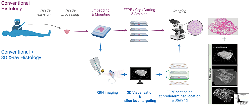
3D X-ray histology (XRH) is a µCT -based workflow tailored to fit seamlessly into current histology workflows in biomedical and pre-clinical research, as well as clinical histopathology.
Non-destructive 3D XRH imaging can be seamlessly integrated into the protocols used for conventional 2D histology and enhance them by providing high-resolution 3D data. The XRH data can also be used to optimise physical sectioning of the tissue block for downstream conventional histology by identifying the areas of interest within the block and slicing accordingly.
Our approach
How we support your project
We welcome collaboration proposals from scientists who want to explore and discover the potential of XRH for their research portfolio. We aim to promote 3D X-ray histology by addressing different communities and levels of technology awareness
The XRH team comprises experienced X-ray beam scientists, microscopists, biomedical researchers and pathologists, together with computing and information scientists. The team is uniquely placed to support and develop a broad range of biomedical imaging applications.
Services include:
Image analysis
Sample visualisation and measurement
Correlative imaging
Combining XRH with conventional histology, immunohistochemistry, and/or other microscopy techniques such as spatial -omics or serial block face SEM
Bespoke analysis
Image base model generation (meshing) for FE/CFD modeling or 3D printing

